Carotid Artery Ultrasound: Accuracy, Duration, and Understanding Color Doppler Imaging
A carotid artery ultrasound is a safe, non-invasive imaging test used to assess the health of your carotid arteries, which are responsible for delivering oxygen-rich blood to your brain. This diagnostic procedure is crucial in detecting narrowing (stenosis), blockages, or other abnormalities that could lead to serious health complications, including strokes.
In this guide, we will address common questions such as:
How accurate is carotid artery ultrasound?
How long does a carotid ultrasound take?
What do the colors mean in a carotid artery ultrasound?
By understanding these aspects, you can gain insight into how carotid ultrasound works, what to expect during the exam, and how it can help in the early detection of cardiovascular conditions.
Get in touch with us!
How Accurate Is Carotid Artery Ultrasound?
Carotid artery ultrasound is considered a highly accurate and effective diagnostic tool for assessing blood flow and detecting vascular disease. When performed by a skilled sonographer and interpreted by an experienced physician, the test provides a sensitivity of over 90% for detecting significant carotid artery narrowing.
Factors Affecting Accuracy
While carotid ultrasound is highly reliable, certain factors can influence its accuracy, including:
- Technician Experience – The expertise of the ultrasound technician and interpreting physician plays a crucial role in obtaining clear images and making precise diagnoses.
- Patient Anatomy – Individuals with short or thick necks may have more challenging scans, making it harder to get clear images.
- Calcified Plaque – Large amounts of calcification in the arteries may obscure the ultrasound waves, leading to limitations in visualization.
- Motion Artifacts – If a patient moves during the scan, it can affect image quality and interpretation.
Despite these variables, carotid ultrasound remains one of the best non-invasive methods for evaluating carotid artery disease, often used as a first-line diagnostic tool before recommending further tests, such as CT angiography or magnetic resonance angiography (MRA).
How Long Does a Carotid Ultrasound Take?
A carotid ultrasound is a quick and painless procedure that usually takes between 15 and 30 minutes to complete. The test requires no special preparation, though patients may be asked to avoid wearing turtlenecks or jewelry around the neck to allow for better access to the carotid arteries.
Step-by-Step Process of a Carotid Ultrasound
1. Patient Positioning
You will lie on your back on an examination table, and a sonographer will apply a warm, water-based gel to your neck.
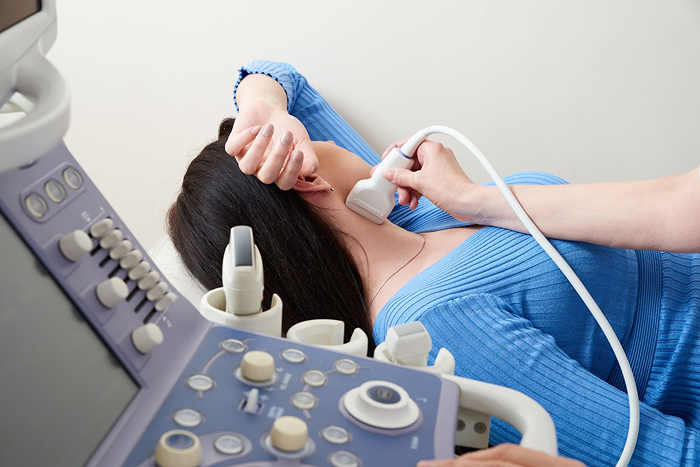
2. Transducer Placement
A small handheld device called a transducer will be moved along the sides of your neck to capture real-time images of your carotid arteries.
3. Image Acquisition
The ultrasound machine will create images using high-frequency sound waves that bounce off the arteries and surrounding tissues.
4. Color Doppler Imaging
To assess blood flow, color Doppler technology will be used to highlight the movement and velocity of blood within the arteries.
5. Completion and Review
Once the exam is complete, the sonographer will wipe off the gel, and your physician will review the results to determine if further evaluation is needed.
Since there is no radiation, no contrast dye, and no needles involved, a carotid ultrasound is completely non-invasive and has no known risks or side effects.
What Do the Colors Mean in a Carotid Artery Ultrasound?
One of the most important aspects of a carotid ultrasound is color Doppler imaging, which helps visualize blood flow within the arteries. This technique allows physicians to evaluate the direction, speed, and patterns of blood flow, which are critical for detecting potential blockages or abnormalities.
Understanding Color Doppler Imaging in a Carotid Ultrasound
-
Red and Blue Colors – These colors indicate the direction of blood flow relative to the ultrasound probe.
- Red typically represents blood moving toward the probe.
- Blue generally signifies blood flowing away from the probe.
- Lighter Shades (Yellow, Orange, Light Blue) – These colors indicate higher velocity or turbulence, which may suggest areas of narrowing (stenosis) in the artery.
- Dark or No Color Flow – If an area appears dark or lacks color flow, it could indicate a severe blockage (occlusion) or very slow-moving blood.
By analyzing these colors, medical professionals can determine whether blood is flowing normally or if there is a risk of reduced circulation due to plaque buildup or other vascular issues.
Why You Should Get a Carotid Ultrasound
A carotid ultrasound is an essential screening tool for individuals at higher risk of stroke or cardiovascular disease. You may benefit from a carotid ultrasound if you have:
A history of stroke or mini-stroke (TIA)
High blood pressure
High cholesterol levels
Diabetes
A family history of carotid artery disease or stroke
Atherosclerosis (hardening of the arteries)
A history of smoking
Early Detection Can Save Lives
Detecting carotid artery disease before symptoms appear can help prevent serious complications, such as stroke. If your ultrasound results show significant narrowing, your doctor may recommend lifestyle changes, medications, or procedures to improve blood flow and reduce your risk of vascular events.
Schedule Your Carotid Ultrasound at Fairbanks Ultrasound
At Fairbanks Ultrasound, we offer state-of-the-art carotid artery ultrasound exams with high accuracy, fast results, and expert interpretation. Our experienced team is dedicated to helping you monitor your vascular health and detect potential issues early.
Do you need help right away?
No problem! Call Us Now.
Fairbanks Ultrasound is a local center in Fairbanks, Alaska that offers various ultrasound services for pregnancy, gynecology, thyroid, vascular, and general purposes.