Edit Content
PCOS Ovary Ultrasound vs Normal
Polycystic Ovary Syndrome (PCOS) is a common hormonal disorder that affects many women of reproductive age. One of the primary diagnostic tools for PCOS is ultrasound imaging, which helps doctors assess ovarian structure and follicle count. But what is the difference between a PCOS ovary ultrasound vs normal? Understanding these differences can provide insight into how PCOS affects the ovaries and how it is diagnosed. By analyzing ovarian size, follicle distribution, and overall structure, ultrasound plays a key role in identifying patterns consistent with PCOS.
What is PCOS?
PCOS is a condition characterized by hormonal imbalances, irregular menstrual cycles, and the presence of multiple small follicles on the ovaries. It can lead to symptoms such as weight gain, acne, excessive hair growth, and fertility issues. Women with PCOS often experience difficulty with ovulation, which can affect their ability to conceive. While blood tests can help assess hormone levels, ultrasound imaging is often used as a non-invasive method to evaluate ovarian health. By comparing a PCOS ovary ultrasound vs normal, medical professionals can determine whether the ovaries exhibit the key characteristics of PCOS or if another underlying condition may be causing symptoms.
Get in touch with us!
How PCOS Affects the Ovaries
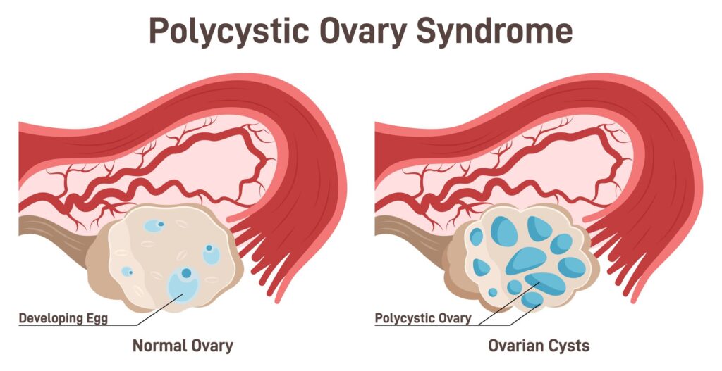
In a typical ultrasound comparison, a PCOS ovary appears different from a normal ovary in several ways. Ovaries affected by PCOS tend to have a higher number of small follicles, which are fluid-filled sacs that contain immature eggs. These follicles fail to develop and release an egg, leading to irregular ovulation or anovulation. In addition to an increased follicle count, PCOS ovaries are often larger in size due to excess follicles accumulating over time. Another defining feature is the “string of pearls” appearance, where multiple small follicles line the periphery of the ovary. This pattern is frequently used by radiologists to distinguish between PCOS and a normal ovarian structure.
PCOS Ovary Ultrasound vs Normal Ovary
A normal ovary, when viewed on an ultrasound, typically has a more uniform and predictable appearance. In women of reproductive age, a normal ovary will contain a moderate number of follicles, with one dominant follicle developing each menstrual cycle in preparation for ovulation. Unlike a PCOS ovary, normal ovaries do not have an excessive number of small follicles, nor do they exhibit the characteristic “string of pearls” formation. Ovarian volume is also a distinguishing factor, as PCOS ovaries are often significantly larger than normal ovaries. Additionally, the stroma, or central ovarian tissue, appears denser in women with PCOS due to increased hormonal activity.

Importance of Ultrasound in Diagnosing PCOS
Ultrasound is a crucial tool for diagnosing PCOS, but it is important to note that an ultrasound alone is not enough to confirm the condition. PCOS is diagnosed using a combination of factors, including menstrual irregularities, clinical symptoms, and hormone levels. The presence of polycystic ovaries on ultrasound is just one criterion and does not necessarily mean a woman has PCOS. Some women may have polycystic-appearing ovaries but do not experience the hormonal imbalances or symptoms associated with PCOS. Likewise, some women with PCOS may have normal-looking ovaries on ultrasound but still meet the diagnostic criteria based on other factors. Because of this variability, doctors use ultrasound in conjunction with blood tests and medical history to provide an accurate diagnosis.
When to Get an Ultrasound for PCOS
Women who experience symptoms such as irregular or absent periods, difficulty conceiving, excessive hair growth, unexplained weight gain, or severe acne may benefit from an ultrasound to assess ovarian health. An ultrasound is particularly useful for evaluating whether ovarian enlargement or an increased follicle count is present. Additionally, women who are undergoing fertility treatments may require ultrasounds to track follicle development and assess ovulation patterns. Doctors may also recommend ultrasounds for women who have metabolic concerns associated with PCOS, such as insulin resistance or an increased risk of type 2 diabetes, as early detection can help with proper management and lifestyle interventions.
Key Differences Between PCOS and Normal Ovaries on Ultrasound
Understanding the key distinctions between PCOS and normal ovaries on ultrasound can help clarify diagnosis and treatment plans. Some of the primary differences include:

Follicle Count and Distribution
PCOS ovaries tend to have an excessive number of small follicles, typically arranged in a "string of pearls" pattern, whereas normal ovaries have a smaller number of well-spaced follicles with one dominant follicle each cycle.
Ovarian Size and Volume
Ovaries affected by PCOS are often enlarged due to the accumulation of immature follicles, whereas normal ovaries maintain a more consistent size throughout the menstrual cycle.
Stromal Density
The ovarian stroma, or central tissue, appears denser in PCOS due to increased androgen production, while in normal ovaries, the stromal area is proportionally smaller and less prominent.
Contact Fairbanks Ultrasound for PCOS Imaging
If you need an ultrasound to assess your ovarian health, Fairbanks Ultrasound offers expert imaging services in a comfortable setting. Our skilled professionals provide accurate ovarian assessments to help guide your diagnosis and treatment. By comparing PCOS ovary ultrasound vs normal, we help patients and healthcare providers make informed decisions about reproductive health and hormone management.
Book an appointment today to learn more about PCOS ovary ultrasound vs normal and take control of your reproductive health.
Do you need help right away?
No problem! Call Us Now.
Fairbanks Ultrasound is a local center in Fairbanks, Alaska that offers various ultrasound services for pregnancy, gynecology, thyroid, vascular, and general purposes.

