PCOS Ovary Ultrasound vs Normal: Understanding Your Ovarian Health
Polycystic Ovary Syndrome (PCOS) is one of the most common hormonal disorders affecting women of reproductive age. Many women experience symptoms such as irregular periods, acne, excessive hair growth, unexplained weight gain, and fertility challenges. A PCOS ovary ultrasound vs normal ovary is one of the most effective ways to evaluate ovarian health and identify structural changes that may indicate PCOS. At Fairbanks Ultrasound, we provide high-quality imaging to help patients understand their reproductive system, confirm diagnoses, and guide treatment planning.
Ultrasound is a non-invasive, safe procedure that uses high-frequency sound waves to create detailed images of the ovaries. By comparing a normal ovary to a PCOS-affected ovary, patients and healthcare providers can better understand hormonal imbalances and potential reproductive concerns. While symptoms and hormone levels are essential for diagnosis, ultrasound provides the visual confirmation needed for a more complete evaluation.
Get in touch with us!
What a Normal Ovary Looks Like on Ultrasound
A normal ovary typically displays a healthy and balanced structure. Follicles, which are small fluid-filled sacs containing developing eggs, change size throughout the menstrual cycle. Normally, one follicle becomes dominant and matures for ovulation, which is an important indicator of healthy ovarian function.
A normal ovary also has a smooth outer surface and uniform internal tissue (stroma). These features indicate proper hormonal balance and ovarian health. On ultrasound, doctors look for subtle changes in follicle size and distribution to ensure the ovary is functioning properly.
Normal Ovary Features
Follicle count is usually between 3–10
Follicles vary in size depending on the menstrual cycle
Follicles are naturally scattered within the ovary
Ovarian volume remains within normal limits (under 10 cm³)
Stromal tissue appears uniform without thickening
A dominant follicle is visible during ovulation
These characteristics help clinicians confirm that ovulation is occurring regularly and that the ovaries are healthy, which is crucial for menstrual regulation and fertility.
What a PCOS Ovary Looks Like on Ultrasound
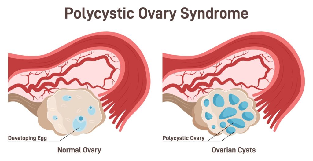
A PCOS ovary shows significant differences compared to a normal ovary. Hormonal imbalances prevent proper follicle maturation, leading to multiple small follicles that remain underdeveloped. These are often mistakenly referred to as “cysts,” but they are actually immature follicles that did not progress to ovulation.
On ultrasound, a PCOS ovary is typically larger than normal and has increased stromal density. The follicles often line the outer edge of the ovary in what is called the “string of pearls” pattern. This distinctive appearance is one of the hallmark signs of PCOS.

PCOS Ovary Features
12 or more small follicles, often more than 20 in some cases
Follicles measure approximately 2–9 mm in diameter
Follicles are arranged along the ovarian perimeter (“string of pearls”)
Ovarian volume is usually increased, often exceeding 10 cm³
Stromal tissue appears denser and brighter on ultrasound
Lack of a dominant follicle due to irregular or absent ovulation
These features help explain the common symptoms associated with PCOS, including irregular or absent periods, hormonal imbalance, and infertility. Ultrasound imaging allows doctors to visualize these changes directly, supporting accurate diagnosis and personalized treatment planning.
PCOS Ovary Ultrasound vs Normal: Key Differences
Comparing a PCOS ovary ultrasound vs normal ovary highlights several important differences. Normal ovaries have a small number of follicles of varying sizes and a smooth, uniform texture. PCOS ovaries have many small, evenly sized follicles along the outer edge and increased stromal density. Enlarged ovarian volume in PCOS contributes to hormonal irregularities and reproductive challenges.
Ultrasound imaging provides clarity for patients, showing why certain symptoms occur and helping guide decisions about fertility treatment, hormone management, or lifestyle interventions. Even in cases where PCOS symptoms exist without classic ultrasound findings, imaging remains an essential component of evaluation.
Why Fairbanks Ultrasound is Your Trusted Choice
At Fairbanks Ultrasound, our team combines advanced technology with compassionate care to provide accurate imaging for women concerned about PCOS or ovarian health. Our skilled sonographers focus on capturing high-resolution images that allow for careful measurement of follicles, ovarian volume, and stromal patterns. We provide patients and physicians with the detailed information necessary to make informed decisions about diagnosis, treatment, and long-term health management.
We understand that reproductive health concerns can be stressful, and we are committed to making every ultrasound experience as comfortable and informative as possible.
Schedule Your PCOS Ovary Ultrasound Today
If you are experiencing symptoms related to PCOS or want to understand the difference between a PCOS ovary ultrasound vs normal, the team at Fairbanks Ultrasound is here to help. Our thorough and compassionate approach ensures that you receive precise imaging, detailed explanations, and support for the next steps in your care.
Contact Fairbanks Ultrasound today to schedule your ovarian ultrasound and gain clarity on your reproductive health.