
MRI vs. Ultrasound: What’s the Difference?
Reviewed by Fairbanks Ultrasound Team
MRI and ultrasound are both painless, non-invasive ways to see inside the body, but they work in fundamentally different ways and are suited to different situations. Ultrasound uses high-frequency sound waves to produce real-time images almost instantly, while MRI uses a powerful magnetic field and radio waves to produce highly detailed, static cross-sectional images over a longer scan time. Neither exam uses ionizing radiation, but the two technologies serve different diagnostic purposes, and your provider will typically choose based on what they’re trying to evaluate, how quickly they need an answer, and your individual health history.
If you live in Fairbanks, Alaska and your provider has ordered imaging, understanding the difference between these two options can help you know what to expect — and in many cases, help you understand why ultrasound was chosen as the first step.
What Is an Ultrasound?
Ultrasound imaging uses a handheld transducer to send high-frequency sound waves into the body. As the waves bounce off different tissues, they generate echoes that a computer converts into real-time images. Because the images appear instantly, ultrasound is especially useful for evaluating movement — such as blood flow through a vessel, the beating of a heart, or a baby’s movement during pregnancy. Ultrasound is widely available, relatively inexpensive, and doesn’t require the patient to enter an enclosed space. At Fairbanks Ultrasound, most exams take 20 to 30 minutes, and same-day walk-in appointments are available with a valid referral.
Get in touch with us!
What Is an MRI?
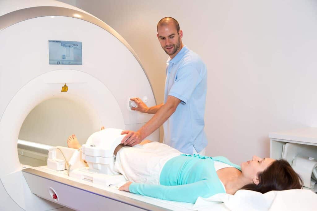
MRI, or Magnetic Resonance Imaging, uses a strong magnetic field combined with radio waves to generate highly detailed, static cross-sectional images of the body’s internal structures. It’s particularly well suited to evaluating the brain, spinal cord, and complex joint or soft tissue detail that ultrasound and CT can’t always capture with the same precision. MRI exams typically take longer than ultrasound — often 30 to 60 minutes — and require the patient to lie still inside a scanner, which can be uncomfortable for patients who experience claustrophobia. Some MRI exams also require an injected contrast agent to enhance certain structures.
Key Differences at a Glance
Several practical factors typically separate the two exams:
Speed: Ultrasound is faster and often available same-day; MRI usually requires more scheduling lead time and a longer appointment.
Cost: Ultrasound is generally significantly less expensive than MRI, both for the exam itself and for patients paying out of pocket.
Real-time imaging: Ultrasound shows movement — like blood flow or a heartbeat — as it happens; MRI produces static images captured over a longer period.
Best for: Ultrasound excels at soft tissue, blood flow, and pregnancy imaging. MRI excels at detailed brain, spine, and complex joint imaging.
Comfort: Ultrasound involves no enclosed space; MRI requires lying still inside a scanner, which can be difficult for patients with claustrophobia.
Equipment access: Ultrasound equipment is widely available in outpatient settings; MRI typically requires a hospital or dedicated imaging center with specialized equipment.
Safety Considerations
Both exams are considered very safe, but each has its own set of considerations. Ultrasound has no known risks and is safe to use during pregnancy, which is one reason it’s the standard imaging method for monitoring a developing baby. MRI, on the other hand, uses a strong magnet, so patients with certain metal implants, pacemakers, or other electronic medical devices may not be candidates for MRI without additional precautions. Your provider will screen for these factors before recommending an MRI.
Can Ultrasound and MRI Be Used Together?
In many cases, yes. A provider might start with an ultrasound because it’s fast and low-cost, and then order an MRI if the ultrasound findings need further clarification or if a more detailed look is required. The two exams are often complementary rather than competing options, each contributing a different piece of the diagnostic picture.
For a similar comparison with another common imaging option, see our related posts:
Why Choose Fairbanks Ultrasound for Your Ultrasound Exam
Fairbanks Ultrasound is a locally owned and operated imaging center specializing in diagnostic ultrasound. If your provider has ordered an ultrasound exam — whether for abdominal, pelvic, vascular, thyroid, or prenatal imaging — we offer same-day walk-in availability with a valid referral, so you’re not left waiting weeks to get the answers you need.
Frequently Asked Questions
For many conditions — especially soft tissue, blood flow, and pregnancy-related imaging — ultrasound is highly accurate and is often the first choice. MRI offers greater detail for certain brain, spine, and complex joint conditions.
Yes, ultrasound is generally significantly less expensive than an MRI exam.
Fairbanks Ultrasound specializes in diagnostic ultrasound imaging. If your provider has ordered an MRI, they can direct you to a local imaging center equipped for that exam.
Ultrasound is typically faster, both to schedule and to complete, with most exams taking 20 to 30 minutes compared to 30 to 60 minutes or more for MRI.
Yes — ultrasound uses sound waves, not magnetic fields, so it doesn’t carry the same restrictions as MRI for patients with implanted medical devices.