
Normal vs. Abnormal Liver Ultrasound: What It Means
Reviewed by Fairbanks Ultrasound Team
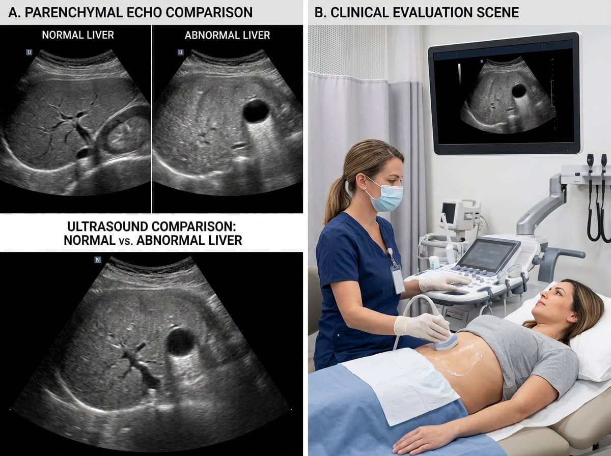
A normal liver ultrasound shows a liver of normal size with smooth edges and an even, uniform texture. An abnormal result usually means the liver looks brighter, rougher, or larger than expected, or that a spot such as a cyst was found. Many abnormal findings, like fatty liver or a simple cyst, are common and manageable. Your provider will explain what your report means and whether you need more tests.
If you are preparing for your exam, start with our guide to liver ultrasound in Fairbanks, AK. If you already have your report, this guide walks through what radiologists look for and what common findings mean.
What Does a Normal Liver Look Like on Ultrasound?
On ultrasound, a healthy liver appears as a large, even-textured gray organ in the upper right abdomen. The radiologist checks several features to decide whether it looks normal:
Size: In most adults, the liver measures about 16 cm or less from top to bottom along the right side.
Surface: The edges are smooth, not bumpy or wavy.
Texture (echotexture): The tissue looks fine and uniform throughout.
Brightness (echogenicity): The liver is about as bright as, or slightly brighter than, the right kidney.
Blood vessels and bile ducts: Vessels are clearly outlined, and bile ducts are not widened.
Blood flow: On a color Doppler view, blood moves in the expected direction through the liver's veins.
Get in touch with us!
If these features look as expected, the report will usually describe the liver as “unremarkable” or “within normal limits.”
Normal vs. Abnormal Liver Ultrasound at a Glance
| Feature | Normal | Possibly abnormal |
|---|---|---|
| Size | About 16 cm or less in most adults | Enlarged (hepatomegaly) or unusually small |
| Surface | Smooth | Nodular or bumpy |
| Texture | Fine and even | Coarse or uneven (heterogeneous) |
| Brightness | Similar to or slightly brighter than the kidney | Much brighter than the kidney |
| Spots or masses | None | Cyst, hemangioma, or other lesion |
| Bile ducts | Normal width | Widened (dilated) |
| Fluid around the liver | None | Free fluid (ascites) |
What Counts as an Abnormal Liver Ultrasound?
An “abnormal” result simply means something differs from the typical appearance. The finding may be minor or may need follow-up. These are the most common abnormal findings.
A Brighter-Than-Normal Liver
A liver that looks much brighter than the kidney usually points to extra fat stored in the liver. This is the most common abnormal liver finding. Learn more in our guide to fatty liver on ultrasound.
A Coarse Texture or Bumpy Surface
A coarse, uneven texture or a nodular surface can be a sign of long-term scarring. Radiologists look at these features closely when evaluating for cirrhosis. See can a liver ultrasound detect cirrhosis for more detail.
Changes in the Bile Ducts or Blood Flow
Widened bile ducts can mean something is slowing bile drainage, such as a gallstone. Color Doppler shows blood flow in red and blue, and changes in flow can point to pressure problems in the liver’s veins. Our guide to what the colors mean on an ultrasound explains how to read those images.
Should I Worry About My Liver Ultrasound Results?
Most abnormal liver findings are common and manageable. Fatty liver is estimated to affect about one in three adults in the United States, and simple cysts and hemangiomas are usually harmless. An abnormal result is a starting point for a conversation with your provider, not a diagnosis on its own.
Contact your provider promptly if you have yellowing of the skin or eyes, severe abdominal pain, vomiting blood, or confusion. Those symptoms need care right away, whatever your ultrasound shows.
What Happens After an Abnormal Liver Ultrasound?
Your provider will combine your ultrasound with your symptoms, history, and lab work. Common next steps include:
- Blood tests such as a liver panel to see how well the liver is working.
- Follow-up ultrasound in a few months to check whether a finding has changed.
- Additional imaging such as CT or MRI to look more closely at a spot. Compare options in MRI vs. ultrasound and CT scan vs. ultrasound.
- A referral to a liver or digestive specialist when more evaluation is needed.
How Long Do Liver Ultrasound Results Take?
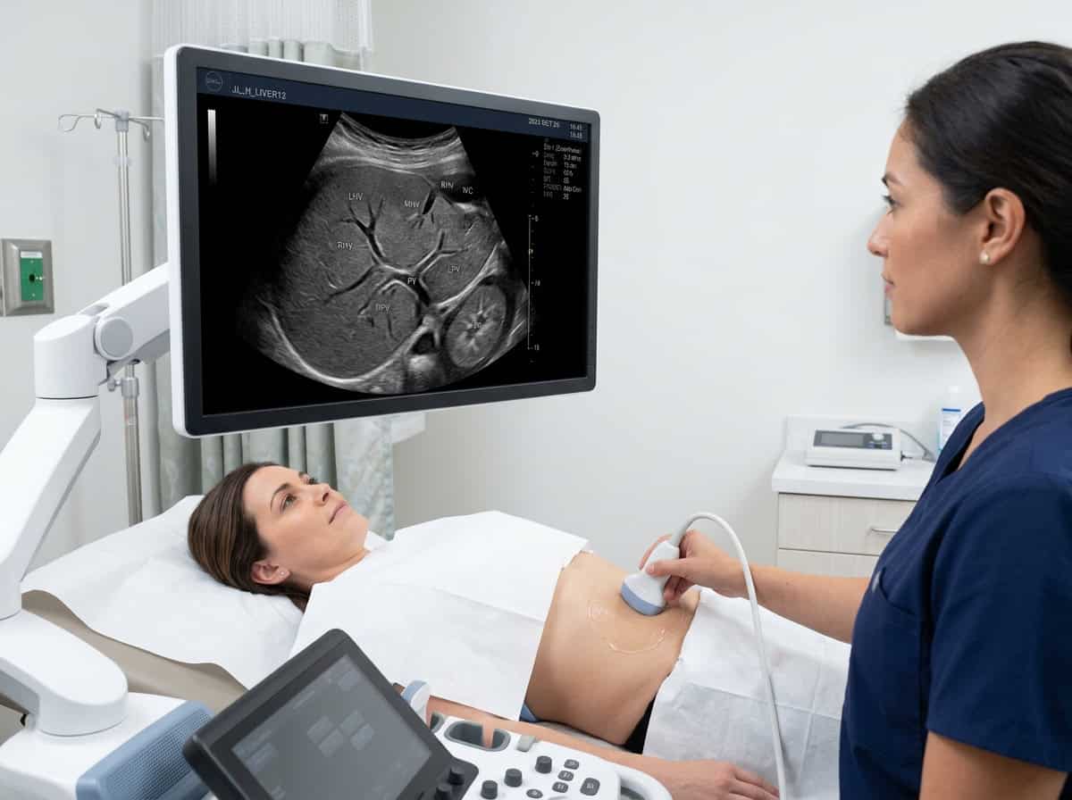
After your exam, a board-certified radiologist reviews the images and sends a report to the provider who ordered the test. The sonographer usually cannot share results during the scan. See how long it takes to get ultrasound results for typical timelines.
Getting a Liver Ultrasound in Fairbanks, AK
Fairbanks Ultrasound is a locally owned imaging center at 570 Riverstone Way, Unit #2, in Fairbanks, Alaska. Our credentialed sonographers perform liver ultrasounds and full abdominal ultrasounds, and our radiologists interpret every exam.
- Follow our fasting prep instructions before your scan.
- Ask about same-day ultrasound appointments with a valid referral.
- Paying out of pocket? Review our ultrasound cost without insurance.
Call (907) 374-4644 or request an appointment. We are open Monday through Friday, 8:00 am to 5:00 pm.
Frequently Asked Questions
A normal liver has smooth edges, an even gray texture, and a normal size. It is about as bright as, or slightly brighter than, the right kidney, and its blood vessels and bile ducts are clearly visible.
Fatty liver is the most common abnormal finding. It makes the liver look brighter than normal and is estimated to affect about one in three adults in the United States.
Ultrasound can show spots or masses in the liver, but it cannot always tell whether a spot is cancer. Most spots are benign. When a finding is unclear, providers usually order a CT, MRI, or other tests.
Usually not. A radiologist reviews your images after the exam and sends a report to your provider, who will explain the results.
Yes, fasting is usually required because it helps produce clearer images. Follow the fasting instructions you receive when you schedule.