
Common Abnormal Findings and What They May Mean
Kidney Stones
Stones usually look like bright spots that cast a dark shadow behind them. Ultrasound is a common first test for stones because it uses no radiation, although very small stones can be missed. CT may be used when a stone’s exact size or location matters. Learn more in can you see a kidney stone on an ultrasound and how to diagnose a kidney stone in Fairbanks, Alaska.
Hydronephrosis
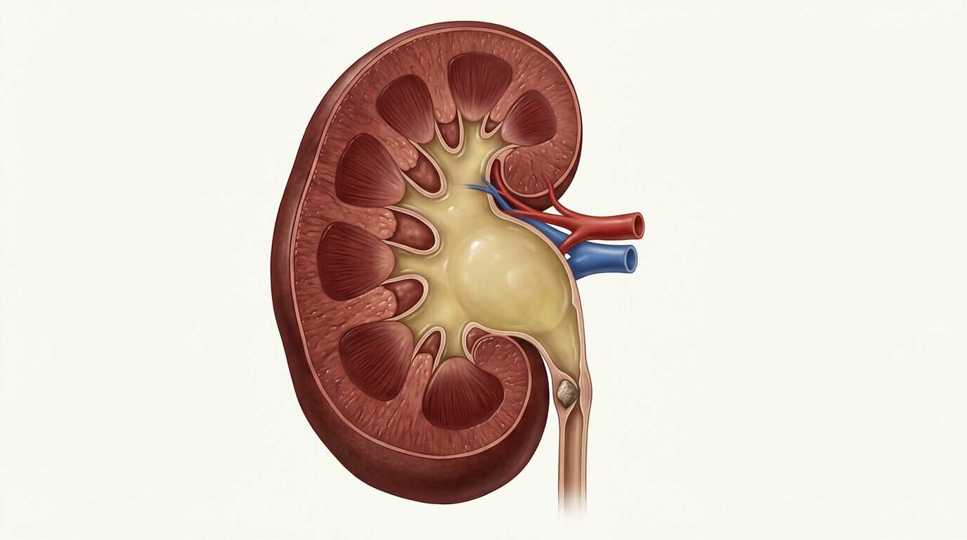
Hydronephrosis means urine is backing up and swelling the center of the kidney. Radiologists often grade it as mild, moderate, or severe. Common causes include kidney stones, an enlarged prostate, and narrowing of the urinary tract. Mild swelling can also appear during pregnancy, and a very full bladder can cause temporary widening, so the sonographer may rescan after you empty your bladder.
Kidney Cysts
Simple kidney cysts become more common with age. By some estimates, about half of adults 50 and older have at least one. A simple cyst is round, fluid-filled, and thin-walled, and it usually needs no treatment. A complex cyst has thicker walls, inner dividers, or solid parts. Complex cysts are usually evaluated further with CT or MRI and sorted with a system called the Bosniak classification. Kidneys that are enlarged and filled with many cysts can point to polycystic kidney disease.
Small, Bright, or Thin Kidneys
Kidneys that are smaller than expected, with a thin outer layer that looks brighter than the liver, usually suggest long-term kidney damage. Blood and urine tests are needed to measure how well the kidneys are working. Read can an ultrasound detect kidney damage or kidney failure to learn more.
Enlarged Kidneys
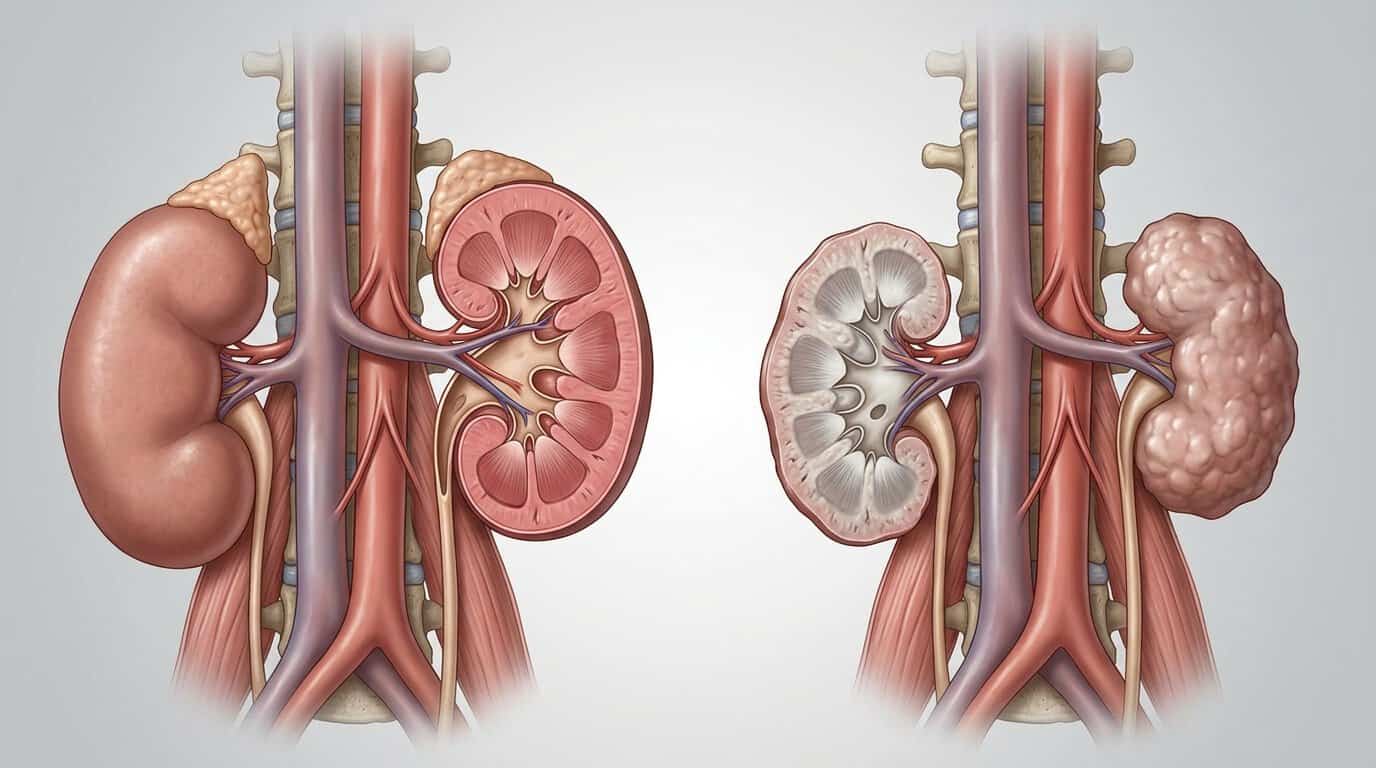
Kidneys that are larger than normal can be linked to polycystic kidney disease, a blockage, infection or inflammation, or some forms of diabetic kidney disease.
Solid Masses
A solid spot in the kidney is not automatically cancer, but it does need a closer look, usually with CT or MRI. Your provider will guide next steps based on the size and appearance of the spot. Read more about what a tumor can look like on ultrasound.
Normal Variations
Some findings are simply differences in anatomy. Examples include a horseshoe kidney (two kidneys joined at the bottom), a duplex collecting system, a column of Bertin, or one kidney that is slightly larger than the other. These are often found by chance and may need no treatment.